The Rhythm of Life: The Beat and Dance of the Heart
Share
- Details
- Text
- Audio
- Downloads
- Extra Reading
The heart beats continuously to sustain life. Its basic rhythm is well known, and has been used as a soundtrack to evoke romance, tension and horror. In this lecture, Professor Elliott will describe how the rhythm is generated. With the help of a percussionist (Nick Buxton of La Shark) and a dancer, he will demonstrate the evocation of those emotions, and what happens when the rhythm of the heart goes awry.
Download Text
12 October 2016
The Rhythm of Life:
The Beat and Dance of the Heart
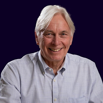
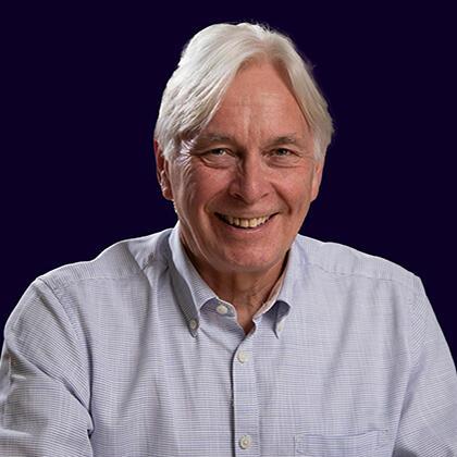
Professor Martin Elliott
“Rhythm is sound in motion.
It is related to the pulse, the heartbeat, the way we breathe.
It rises and falls.
It takes us into ourselves; it takes us out of ourselves.”
Edward Hirsch
Introduction
I want to remind you of the magic of the heart. From shortly after conception and throughout your life, it works ceaselessly. It beats 100,000 times a day; 40 million times a year; or 3 billion times in an average lifespan. Mostly without us being aware of it.
How does that happen? What makes this muscle work on its own, and with such astonishing regularity, reliability, responsiveness and rhythm? Do these mechanisms ever go wrong, and what are the consequences if they do? I am going to try and answer these questions tonight, but in a slightly different way to a conventional lecture.
I am honoured and privileged to have the help of two drummers; Nick Buxton, an artist in wood, and brilliant drummer with the band ‘La Shark’ who will help us tease out some of these complex rhythms. And the actor Hanna Harlyn who will show us in dance how these rhythms affect the heart.
First though, there is some biology to get out of the way.
Our bodies contain three basic types of muscle, smooth muscle (found for example in the gut), striated or skeletal muscle (the ones we see in the gym) and cardiac muscle. Whilst these muscle types share some structural and functional characteristics, there are significant differences.
Within the heart, cardiac muscle cells take up about 2/3 of the structural space available, but make up only about a third of the cells. The other two thirds of cells (fibroblasts, blood vessel cells, neuronal and inflammatory cells) comprise the extracellular matrix and the blood vessel system of the heart which are not only structural, but provide both macro- and micro-anatomic barriers that are central to the electrical function of the heart, known as electrophysiology1.
The main heart muscle cell is the cardiomyocyte. These are rod-shaped cells roughly 100 μ (about the diameter of a human hair) by 20μ. They are wrapped around by an insulating lipid bi-layer 80 – 100Å thick. This layer has an important role in managing the electrical functions of the heart. The cells form a branching network, joined together at intercalating discs bound by a sort of inter-cellular Velcro called desmosomes. There are also electrically important connections between adjacent cells called gap junctions, critical to the rapid propagation of electrical signals over many cells.
Here is a brilliant nano-movie of a mouse heart cell contracting2 .The intracellular myofibrils contain actin and myosin molecules which can be thought of as sliding over each other3. The myofibrils are organised into a chain of contractile units called sarcomeres. These are what gives the muscle its banded appearance.
This animation (https://www.youtube.com/watch?v=hr1M4SaF1D4) shows the primary process of contraction in muscle. Myosin is a thicker filament than actin. Myosin is anchored at the central M-line of the sarcomere, and actin is anchored at the end, or Z-plates. This means that when the actin filaments slide along the myosin, the sarcomeres shorten from both ends. This happens synchronously so the whole muscle shortens.
Although it is conceived as a sliding movement, in fact the myosin pulls the actin over its length by forming cross-bridges at binding sites on the actin molecule. Bound ATP is hydrolysed to ADP and Pi which causes the myosin filaments to extend and attach to a cross bridge. An action called the power stroke is triggered causing actin to move towards the M-line, thereby shortening the sarcomere and contracting the muscle. ADP and Pi are released during the power stroke and the myosin remains attached to the actin until a new molecule of ATP binds, feeing the myosin to go through another cycle of binding and more contraction or remain unattached to allow the muscle to relax.
Muscle contractions are controlled by the actions of calcium. The thin molecules of actin contain regulatory proteins called troponin and tropomyosin. In the relaxed state, tropomyosin blocks the binding receptor sites on the actin molecule. When calcium ion levels are in high enough concentration, and enough ATP is present, calcium ions bind to the troponin, which displaces tropomyosin exposing the myosin binding sites on the actin allowing myosin to form cross-bridges and continue the cycle of contraction and relaxation. Contraction and relaxation of muscle thus require chemical energy, and are crucially dependant on calcium ions.
What Causes the Muscle to Start to Contract.
Some form of electrical signal is necessary to trigger contraction of the muscle. To explain this, I need briefly to describe the importance of the cell membrane in the process of propagating contraction. The insulating lipid bilayer on the outside of the cardiac muscle cells permits little transport of ions and maintains a separation of charge established by active transporters that reside in the cell membrane. Electrically charged ions, particularly of [NA], [K] and [Ca] exist in different concentrations on either side of the cell membrane, maintaining a polarised state across the membrane, with the inside being –ve in relationship to the outside. Thus there is a r resting membrane potential, measured in mV. This membrane potential has to be maintained by an active process driven by electrogenic pumps (Na+/K+ ATPase and Ca++ transport pumps). Ion channels are transmembrane proteins that serve as a conductive pathway between the inside and outside of the cell, allowing the flow of ions and thus charge (i.e. current).
When the cell is appropriately stimulated, the negative voltage inside the cell may transiently become positive owing to the generation of an action potential. The action potential is basic to the process of stimulating contraction of muscle, and it can spread across muscle as there is current flow among the myocytes. The heart cells are connected to each other by low resistance communications called gap junctions which contain intracellular ion channels4 whose porosity to ions can be varied.
The processes involved in generating the action potential are extremely complicated, and I am grateful to the team at Bio VFX for this simplified explanation, which I think works quite well. [please watch the on-line video at this point] This red box represents a cardiac muscle cell. Within that cell, you can see a small amount of sodium, represented by the salt in this salt cellar, a small amount of calcium represented by this bone, and a large amount of potassium represented by this banana. The amount of each electrolyte is represented by how full each item is. The potassium is 90% full, and the sodium and calcium are only about 10% full.
Outside the cell are the same three electrolytes, potassium, calcium and sodium. Outside the cell, potassium is only 10% full, but sodium and calcium are 90% full. All these electrolytes have a positive charge, so there is more positive charge on the outside of the cell and the inside is less positive, i.e. negative. What…I hear you say?!! Let’s imagine that we can count the amount of positive ions in our diagram. On the outside we have 90 sodium + 90 calcium + 10 potassium equalling 190 + charges outside the cell. On the inside, we have 10 sodium + 10 calcium + 90 potassium giving us a total of 110. Thus the outside of the cell is more positive than the inside. When these ions start to move, this is what happens.
Sodium moves slowly into the cell, and then rapidly causing potassium to exit the cell. The exit of potassium causes calcium to enter the cell, change the concentration of all the ions, and the inside of the cell becomes more positive than the outside. The movement of the ions has caused a voltage change across the membrane.
This can be shown graphically. This horizontal time represents time, and the vertical line represents millivolts. As sodium moves slowly into the cell, there is a gradual change in the voltage until the transmembrane potential reaches about -70 mV, at that point a threshold is reached, and sodium rushes in and potassium rushes out and the inside of the cell becomes frankly +ve. The loss of potassium causes the voltage to drop a little and then, as the calcium flows in, things equalise out and there is little change in voltage. This is called a plateau. The cell now starts actively to pump sodium out and potassium in, and at the same time calcium leaks out of the cell. The transmembrane potential becomes more negative again, eventually reaching a steady state of the resting membrane potential….this horizontal line. These events happen with every heartbeat.
The conduction of electricity through the heart is very organised, resulting in maximal efficiency of the heart as a synchronous and responsive pump. To the humble surgeon, it seems like magic.
But where and how does the heart beat start?
The beat of the heart begins in a group of specialised cells, sometimes called pacemaker cells, (discovered by Martin Flack in 1907), which have a particular set of processes which make them depolarise automatically, at the fastest natural rate of the heart, and able to be influenced by nervous pathways and hormones.
A great deal is now known about these pacemaker cells, but the key feature of them is that they do not have to wait for a stimulus to depolarise, they do so automatically. Instead of a flat line of the resting potential during the resting phase, diastole, of the heart, pacemaker cells gradually depolarise automatically, as shown in this slide. The automatic depolarisation of the pacemaker cells occurs as a result of timed ion exchanges4 controlled by what have been termed ‘clocks’ at membrane level and related to the release of calcium from part of the cell called the sarcoplasmic reticulum5. This creates cycles of activity which have been defined as ignition, depolarisation, resetting, refuelling and repolarisation, as seen in this diagram5. It is way too complex to describe here (although this diagram gives a flavour5), but offers innumerable sites for drugs to modify conduction, and thus to treat abnormal rhythms.
The electrical signal begun by the pacemaker cells, then spreads through a system in the heart called the conduction system. This animation [see accompanying video of lecture], made by Alila Medical Images (http://www.alilamedicalmedia.com), shows how the events of the conduction system relate anatomically to what is seen in the electrocardiogram (ECG) recorded via electrodes on the skin. This ECG recording is the summation of the action potentials of the heart.
The beat of the heart begins in the pacemaker cells of the sino-atrial node, situated near where the superior caval vein enters the right atrium. It spreads rapidly across the atrium from right to left, as the P-wave on the ECG causing the atria to contract and squeeze blood into the empty ventricles through the tricuspid and mitral valves.
The atria and ventricles are separated from each other by an effective insulating barrier, except for a specialised conduction area called the atrio-ventricular node which briefly delays the flow of the signal before passing it down the bundle of His. The bundle of His splits into two branches in the interventricular septum: the left bundle branch and the right bundle branch; both head off towards the apex of the heart. The left bundle branch activates the left ventricle, while the right bundle branch activates the right ventricle. The left bundle branch is short, and itself splits into two fascicles. The left posterior fascicle is relatively short and broad, with dual blood supply, making it particularly resistant to ischemic damage. As the left posterior fascicle is shorter and broader, impulses reach the papillary muscles just prior to depolarization, and therefore contraction, of the left ventricle myocardium. This allows pre-tensioning of the chordae tendinae, increasing the resistance to flow through the mitral valve during left ventricular contraction. This mechanism works in the same manner as pre-tensioning of car seatbelts.
The passage of this current along the bundle and fibres of the ventricles corresponds to the QRS complex on the ECG. This rapid conduction pathway, combined with the slight slowing down of the signal at the AV Node, means that atria can empty completely into the ventricles before the ventricles themselves start to contract, which they do from the apex up towards the aorta and pulmonary artery, the two exit routes. This atrio-ventricular delay is critical to prevent inefficient filling, and ensure appropriately timed valve closure and thus avoid backflow or flow collisions.
Sending the signal to the apex first makes sure the blood is squeezed in the correct direction (like squeezing toothpaste) and increases the efficiency of the whole process. The atria relax and fill at the same time, and the whole process can start again after repolarization of the cells represented by the T wave. Relaxation of the heart is rapid and organised, with temporary inactivation of appropriate ion channels.
In fact, thanks to the wonderful maze of connections between the muscle cells in the ventricles, the depolarisation spreads really quickly, and the heart muscle cells contract almost simultaneously. Compared with other muscle cells, the action potential of the heart cells lasts a relatively long time. This stops the muscles cells from relaxing too soon, keeping contraction going until all the muscle has had time to depolarise and contract as an efficient effective single unit.
To recap, the signal from the pacemaker cells in the sino-atrial node cells is propagated across the atria, the muscle cells of which contract as the signal travels cell to cell via gap junctions creating a sort of integrated unit, squeezing the contents of the atria into the ventricles. This rapid conduction pathway, combined with the slight slowing down of the signal at the AV Node, means that atria can empty completely into the ventricles before the ventricles themselves start to contract, which they do from the apex up towards the aorta and pulmonary artery, the two exit routes. This atrio-ventricular delay is critical to prevent inefficient filling, and ensure appropriately timed valve closure and thus avoid backflow or flow collisions.
Having got that basic stuff out of the way, let’s talk about rhythm and, first, rate.
An impulse (action potential) that originates from the SA node at a relative rate of 60 – 100 bpm is known as normal sinus rhythm.
If SA nodal impulses occur at a rate < 60 bpm, the heart rhythm is known as sinus bradycardia.
If SA nodal impulses occur at > 100 bpm, the consequent rapid heart rate is sinus tachycardia.
Note though that your heart rate varies with age, fitness and activity.
When it all goes wrong
When the rhythm of the heart goes awry it is called an arrhythmia or dysrhythmia. These may be silent, with no symptoms or on occasion the patient will notice something is not right. I will talk about some of these symptoms as we go on.
There are four main types of abnormal rhythm:
- Extra or premature beats
- Supra-ventricular tachycardias [fast]
- Ventricular arrhythmias
- Brady [slow] arrhythmias
Nick, Hanna and I are going to take you through these to describe them. We don’t have time to present all the abnormal rhythms, some of which are extremely rare; it would take too long and miss the point. We want to leave you understanding more, but not everything, about what can happen and what the consequences might be. Otherwise there would be little work for my cardiology colleagues to do!
Extra Beats
Let’s start with extra beats, and first, Premature Atrial Contractions (PACS). These are very common, are normal at any age and may be either completely unnoticed or felt as a skipped beat. Usually that means that one heart beat feels a bit late or maybe a little more prominent. This is an ECG showing PACS. In amongst the normal, regular rhythm of the heart, a beat appears early, followed by a compensatory pause as repolarisation gets back to normal. Let’s watch while Nick and Hanna show you what it sounds and looks like!
They happen when another part of the atrium depolarises before the SA node. Why this happens is not yet fully understood, although they can be more common after caffeine for example. They only need treating if they are causing anxiety or in rare cases with other heart disease when they can trigger more serious atrial rhythm problems. Beta-adrenergic blockers work quite well.
The other extra beat rhythm is Premature ventricular contractions (PVCs). These look very different from PACs on an ECG, but they are quite common and equally benign. They may also be felt as a skipped beat or if a few occur together as palpitations[1]. A PVC starts when a beat is initiated early by Purkinje fibres in the ventricles rather than the ordinary pathway. There is no atrial contraction as you can hear and see now!
There are more important abnormal rhythms which are characterized by their origin and their rate.
Supra-Ventricular Tachycardias (SVT)
These are defined as fast heart rates originating above the AV node. They usually occur in ‘bursts’ and so end up being called paroxysmal SVTs or PSVTs. Because these episodes last longer than just one or two beats, they are more often associated with symptoms, which can be quite alarming. For example, patients may experience a pounding heart, shortness of breath, chest pain, rapid breathing, dizziness and (in very severe cases) loss of consciousness.
There are several types of SVT related to the cause of triggering the heartbeat.
- Sino-atrial node re-entrant tachycardia (SANRT). Re-entry rhythm problems occur when an electrical impulse travels in a tight circle within the heart rather than the conventional pathway, and these occur because there is some variation (heterogeneity) of the refractory period of (especially) thin walled muscle. SANRT usually begins from areas so close to the SA node that ECG looks just like fast normal, unless you see or record the initiation event. The net effect is very like a very fast regular rhythm…too fast sometimes to give the heart time to fill properly between beats, leading to the symptoms. It will sound and look like this. By the way, you can usually slow down by massaging the neck (carotid sinus) or pressing on the eyeball. These trigger the vagus nerve which sends parasympathetic signals to the heart to alter ionic flux.
- Automaticity. In this type of SVT, the onset is usually gradual as a very localised area stimulates the very fast beat without a re-entry pathway.
- Atrial Flutter. This is a really fast regular rhythm of up to 300 bpm in the atria caused by a re-entry circuit (loop of depolarisation) in the atria which overrides the SA node. Atrial flutter is often associated with an underlying disease (like ischaemia) which may make the heart cells irritable. Because the refractory period of the AV node is 330ms the maximum conduction rate is 1:1 at 300bpm, so rates faster than that do not conduct 1:1 but 2:1, 4:1 or rarely 3:1. The pulse rate is regular.
- Junctional Rhythms. There are some specific rhythms which produce flutter-like behaviour of the atria, but because of abnormal re-entry pathways close to the AV junction, hence the name junctional rhythms. There is now a well-established list of potential accessory pathways like this [slide] and it is now possible to both identify and ablate these pathways either with a cryo-probe or with a radiofrequency pulse [movie]
- Atrial Fibrillation This is the most common abnormal heart rhythm, occurring in 2-3% of the population. Instead of the atrial muscle cells beating together in a synchronised way, they contract independently of each other resulting in a disorganised and ineffective contraction which makes the atrium look like a quivering bag of worms, preventing effective ventricular filling. The normal signals from the SA node are completely overwhelmed by disorganised electrical impulses which largely originate close to the pulmonary veins, where oxygenated blood returns to the left atrium from the lungs. Over time, if AF continues, the atrium gradually fibroses or scars as the atria dilate. The scarring can spread to the pacing nodes, making a vicious circle. Because AF discharge rates are so high, most do not end up in a ventricular beat, only getting through occasionally. Thus there is no ‘beat’ of the atria, and the ventricular response is irregularly irregular. As we will now demonstrate……
-
- The incidence rises with age, so almost 14% of the over 80’s have it. Flutter and fibrillation are said to be causes of death in 112,000 deaths in 2013. It often starts with short bursts, but become more prolonged and sometimes constant. Apart from the general symptoms we discussed earlier, there is a risk of heart failure and stroke and some have suggested dementia.
- In only half the cases is some sort of cause identified, ranging from ischemic and valvular heart disease to Chronic Obstructive Pulmonary Disease, excess alcohol intake and many others.
- If at least one of your parents had AF, your chances of getting it are almost doubled compared with the rest of the population, and it is associated with at least 4 types of abnormal genes.
- Diagnosis by ECG, ECHO and a search will be made for underlying, and especially correctible, causes. Treatment is essentially the same as for severe atrial flutter..
Brady (slow) rhythms
If the SA node fails to start its signal for some reason, the AV junction can take over as the main pacemaker of the heart. The AV junction consists of the AV node, the bundle of His and the surrounding area; its natural regular rate is much slower than the native SA node speed (60-100) and has a regular rate of 40 to 60bpm. These "junctional" rhythms are characterized by a missing or inverted P-Wave. If both the SA node and the AV junction fail to initialize the electrical impulse, the ventricular muscle cells take over and fire themselves at a rate of 20 to 40bpm and will have a QRS complex of greater than 120ms.
There are some specific slow heart rates that you will undoubtedly have heard of. These are called heart block, and there are basically three types with Type 2 itself having two variants.;
- First Degree Heart Block, in which there is a delay in conduction between the SA node and the AV node as the signal passes across the atria. This results in a prolongation of the P-R interval on the ECG increasing the gap between atrial contraction and ventricular contraction.
- Second Degree Heart Block, itself made up of 2 main types, Mobius Type 1 (Wenckebach) and Mobius Type II.
-
- In the former, the delay to conduction takes place in the AV nodal area. Some of the AV nodal cells tend to fatigue until they fail to conduct an impulse. This results in a progressive prolongation of the PR interval until the P wave is not conducted to the ventricles, and no ventricular contraction occurs for a beat. The QRS complex is narrow. Let’s show you what that means. It is usually fairly benign.
- In Mobius Type II, which is much more important medically, the P waves continue to occur at a normal rate, but they are only intermittently conducted, resulting in this sort of rhythm. The problem occurs because of some physical abnormality in the Bundle of His or beyond, and the QRS complexes are wide. Ventricular contraction may be relatively inefficient, and with the low rate, the cardiac output is reduced. This often proceeds to become complete or 3rd degree heart block and may indicate a pacemaker insertion.
- Both types may end up producing a fixed-rate 2nd degree heart block in which only affixed proportion of p waves get through to result in a ventricular beat, for example 2:1, which looks and sounds like this. Or 3:1 like this.
- Third Degree Heart Block, in which no P waves get through to the ventricles at all. The P waves just carry on at their own rate, independent of the ventricles, which work at their own, slower, escape rate. The slower the rate, the more dangerous the situation, as simply not enough blood may be ejected to meet the needs of the body, and urgent treatments, in the form of drugs which increase the rate, or more likely a pacemaker, are required.
- Left Bundle Branch Block. You can get block of just one branch of the AV bundle. If this occurs on the left branch, to the left ventricle, the electrical stimulus travelling through the conduction system arrives in the right ventricle first, and then has to spread across to the left, thus delaying left ventricular contraction.
- Right Bundle Branch Block. A similar situation occurs if the right bundle is damaged. Here right ventricular contraction is delayed. And so is Hanna’s right leg!
Fast Ventricular Rhythms If that wasn’t hard enough for Hanna to demonstrate...this is going to be much more exhausting.
- Ventricular Tachycardia (VT). This is a very fast heart beat that starts from inappropriate electrical activity in the ventricles. The longer it lasts, the more dangerous it is and symptoms develop quickly if it goes on longer than 30secs (sustained VT). These symptoms include light-headedness, palpitations or chest pain as the rate out paces the blood supply. Rates exceed 120 bpm, and the pattern on ECG may be monomorphic in which the QRS complexes are uniform and broad on all leads of the ECG, and the signal starts from a single reentry circuit somewhere in the ventricle, usually associated with some area of damage. Essentially, it is what its name suggests…a very fast ventricular rhythm. This rhythm may be quite well tolerated for some time, but the faster the rate (upto 270 bpm), the more dangerous it becomes and the shorter the time you can tolerate it before collapse.
The other variant of ventricular tachycardia called polymorphic (torsade des pointes) is more dangerous. In this type of VT, abnormal triggers for the heart beat start in multiple palaces through the ventricles, and there is quite a significant variation beat to beat. This results in a very low and ineffective cardiac output and can cause collapse and death quite quickly unless reversed by DC shock.
- The last rhythm I want to demonstrate is Ventricular Fibrillation. You will remember that fibrillation is quivering, and in this rhythm all the ventricular cells are beat independently, at random and with no synchrony. The ventricles look like a bag of worms, and there is no forward flow. Without urgent treatment, this is fatal. No blood reaches the vital organs. Fortunately, you can treat it by applying a DC shock across the heart with a defibrillator. You have to see it to believe it. So here goes.
It is easy for us in the operating theatre to sort this out. The ECG shows the problem, and we have all the equipment we need. If someone collapse in the street in front of you, and you don’t have all this stuff available. What can you do? It would be remiss of me not to tell you. You could save a life.
Here Denise Welsby comes on and takes us through what to do to bring Hanna back to life.
Ladies and Gentlemen. Maybe the words of Ringo Starr are apt “Rhythm comes from the body, and timing from the heartbeat. I try to teach this to kids – some get the idea and some don’t”. I hope you leave here with a clearer picture of the abnormal heart rhythms that exist. I hope you appreciate again how amazing it is that the heart just keeps on beating.
Please join me in thanking Hanna Harlyn, Denise Welsby, and Nick Buxton for being so brave.
Thank you.
© Professor Martin Elliott, 2016
References
1. Saksena S, Camm AJ, editors. Electrophysiological Disorders of the Heart. Philadelphia, PA: Elsevier, 2004.
2. Kobirumaki-Shimozawa F, Oyama K, Shimozawa T, et al. Nano-imaging of the beating mouse heart in vivo: Importance of sarcomere dynamics, as opposed to sarcomere length per se, in the regulation of cardiac function. The Journal of General Physiology 2016;147(1):53-62.
3. Cooper GM. Actin, Myosin and Cell Movement. In: Cooper GM, ed. The Cell:A Molecular Approach. Washington: Sinaeur Associates, 2000.
4. Tomaselli G, Roden DM. Molecular and Cellular Basis of Cardiac Electrophysiology. In: Saksena S, Camm AJ, eds. Electrophysiological Disorders of the Heart. Philadelphia, PA: Elsevier, 2004:1-31.
5. Monfredi O, Maltsev VA, Lakatta EG. Modern Concepts of the Origin of the Heartbeat. Physiology 2013;28:74-92.
Special Thanks to
Hana Harlyn
Nick Buxton
Denise Welsby
Becan Rickard-Elliott
Lesley Elliott
Scott Rimington
Dr Jasveer Mangat
Gresham College
Barnard’s Inn Hall
Holborn
London
EC1N 2HH
www.gresham.ac.uk
[1] a perceived abnormality of the heartbeat characterized by awareness of heart muscle contractions in the chest: hard beats, fast beats, irregular beats, and/or pauses.
References
1. Saksena S, Camm AJ, editors. Electrophysiological Disorders of the Heart. Philadelphia, PA: Elsevier, 2004.
2. Kobirumaki-Shimozawa F, Oyama K, Shimozawa T, et al. Nano-imaging of the beating mouse heart in vivo: Importance of sarcomere dynamics, as opposed to sarcomere length per se, in the regulation of cardiac function. The Journal of General Physiology 2016;147(1):53-62.
3. Cooper GM. Actin, Myosin and Cell Movement. In: Cooper GM, ed. The Cell:A Molecular Approach. Washington: Sinaeur Associates, 2000.
4. Tomaselli G, Roden DM. Molecular and Cellular Basis of Cardiac Electrophysiology. In: Saksena S, Camm AJ, eds. Electrophysiological Disorders of the Heart. Philadelphia, PA: Elsevier, 2004:1-31.
5. Monfredi O, Maltsev VA, Lakatta EG. Modern Concepts of the Origin of the Heartbeat. Physiology 2013;28:74-92.
Part of:
This event was on Wed, 12 Oct 2016
Support Gresham
Gresham College has offered an outstanding education to the public free of charge for over 400 years. Today, Gresham College plays an important role in fostering a love of learning and a greater understanding of ourselves and the world around us. Your donation will help to widen our reach and to broaden our audience, allowing more people to benefit from a high-quality education from some of the brightest minds.


 Login
Login


