Mimicry of Nature for Disease: Detection and Destruction
Share
- Details
- Transcript
- Audio
- Downloads
- Extra Reading
'If one way be better than another, that you may be sure is Nature's way' (Aristotle).
Mimicry forms the basis of Medical Biomimetics, the imitation of nature to treat disease.
During the last 3.6 billion years, nature has gone through a process of trial and error to refine processes and materials of life. This emerging field has given rise to new technologies created from biologically inspired engineering, such as using light/rainbow analysis for early detection of disease and using light to clear to cancer, analogous to plant reversal of photosynthesis using photo destruction for autumnal sensecence.
Download Transcript
23 October 2014
Mimicry of nature for disease:
Detection and Destruction
Professor Hugh Barr
‘if one way be better than another, that you be sure is Natures’s way’ Aristotle.
Mimicry forms the basis of Medical Biomimetics - the imitation of the nature to detect and treat disease. During the last 3.6 billion years; nature has gone through a process of trial and error to refine the processes and materials of life. Mimicking nature has given rise to new technologies created from biologically inspired engineering. In particular for non-invasive and none destructive rapid real-time diagnosis of disease: the devices developed are strongly based on nature’s solution.
This lecture will explore the use of smell (odour detection), ‘sniffing out disease’ to ‘snuff it out’: and the use of optical detection (spectroscopy) or ‘rainbow analysis’; for the early detection of cancer. Removing tissue (biopsy) to diagnosis is ‘damaging to detect’. This is impossible in vulnerable areas and is wasteful and unnecessary.
If we use the principles of light harvesting during photosynthesis we can exploit nature’s photo destructive reactions to target and destroy diseased areas. This is utilizing the processes occurring in the cyclical natural, massive and beautiful destruction of leaves during autumnal senescence.
Odour The doctor will smell/see you now!
‘Noses have they, but they smell not’ David’s lament to the people in Psalm 115 v5.
You are blind and deaf worm; how can you interact with the world outside? How can you identify enemies and friends and indeed future partners or mates? Your information is obtained entirely by chemosensory mechanisms (small and taste). The odours that are liberated into the general atmosphere around you are vital to your survival and that of your species. Indeed for many lower order mammals it is also a major window on the external environment. In some mammalian genomes there are over 1000 genes coding for chemosensory and is at least 1% of the entire genome greater than that of the immune system.
Our human sense of smell is in comparison very poor since we have developed complex organs (the eye) as our primary method of environmental and social assessment. This dominance is very evident in our current obsession with interaction via the social media, computer screens and television. This trend is very likely to continue with telemedicine and doctor patient relationships conducted by the telephone or Skype.
Our greatest smell deficiency happened many millennia ago with the loss of a meaningful Vomeronasal organ for social interactions to assess pheromone signals.
This chemosensory ‘Organ of Jacobson’, is vital and well utilized in reptiles and non-primate mammals. It is atrophic and rudimentary in man. Behaviour modification, reproductive, sexual attraction and subsequent sexual behaviour of lower organisms is by the detection in this organ of simple molecules (eight to ten carbon atoms). They are highly specific to certain receptors and very potent drivers of behaviour. For example when a female moth releases ‘Bombykol’ the male will travel great distances at great peril for the promise of an ever so transitory and brief mating. Locust aggregation is driven by Guaiacol released from the faecal pellet, and produced by the insect’s intestinal bacterium.
What of the odours noticed by our noses and also not detectable but interacting with the remnant of our vomernasal organ. These are present in the air around us and all pervasive. The response to some chemicals is to allow social bonding and encouraging of monogamous relationships. Other chemicals may induce a more feral and promiscuous attitude.
Also can your smell be a sign of disease, decay and deterioration. Patients with schizophrenia do release a distinct odour in their sweat based on trans-3-methylhexanoic. May we smell you to help you? All living organisms from complex species to microorganisms produce and excrete by-products as a result of their normal metabolism. The ability of different organisms to metabolize different substrates in order to satisfy their energy and nutritional requirements is fundamental to their life and environmental interaction. Some of these metabolic products, including alcohols, aliphatic acids, ketones and terpens are liberated as volatile organic compounds (VOCs). Many VOCs have characteristic features, but some are not an obvious distinct odour. VOCs production is not limited to microorganisms and volatile fingerprints can be traced in conditions such as cancer, metabolic disorders, mental disorders, and cardiopulmonary disease. Since the production patterns of VOCs are unique to certain micro-organisms or disorders, they can potentially be used as biomarkers to detect disease. Humans are not highly dependent on olfaction since they have to cope with a very diverse range of odour molecules. There are over 1000 odorant receptors in the nasal olfactory bulb. Generally we are overly dependent on vision and hearing to detect diseases. Compared to the rest of the natural world we are deficient in the use of our chemosensory (smell and taste) apparatus.
The nature of odours.
The fragrance of the flower, putrefaction, the odours of decay, the breath of a diabetic patient, and obvious mustiness of a patient entering renal or hepatic failure are complex mixtures of many thousands of volatile chemicals, but not all are meaningful. We are not constantly aware of all odours around us but can tune into specific chemicals. We are able to assign specific identities to mixtures, but precise distinction, unlike the visual system, is not highly specific. The physics of smell are also intriguing. Our eyes look at specific areas and our hearing can also localise position well. Volatile agents waft on the breeze and are part of our turbulent environment. Indeed, many will only be detected by ‘sniffing’, as occurs with a bee in a flower or more commonly a dog detecting smell on a tree; or the perineum of another dog. Thus odour sampling has to occur and most mammalian species sniff, and insects move their olfactory appendages. This behaviour controls the sensory input in time and space since olfaction is poor at following very rapidly varying signals.
Smell and Disease.
The foul smell associated with bacterial infection and rotting flesh has long been recognised. The Miasmatic theory suggested that many diseases were caused through the emanations arising from the corruption infecting the air. There were severe punishments for ‘those which cause corruptions near a city to corrupt the air’. Great efforts were made to create healthy odours; forming the basis of the perfume industry to enhance attraction.
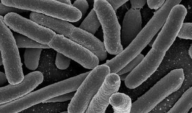
It is now clear that many bacterial species produce specific odours, most notably the anaerobic bacteria such as Bacteroides and Clostridia species. Often a pungent odour can only be detected if there is an overwhelming infection. Yet, mild infection in the urine of a child with Eschericha Coli imparts a distinct fishy odour, often detected by the mother familiar with the normal smell.
Certain metabolic diseases may be first detected on the patient’s odour. Prior to the advent of molecular and biochemical medicine, the sweet smell imparted to the breath of an unconscious child was the first clue of an acidotic diabetic coma. A musty odour in an older patient could indicate imminent hepatic failure, or a urinose smell, kidney failure. Halitosis or simple bad breath may have many causes, but is often related to excessive bacterial overgrowth in the stomach associated with gastric cancer or in the lungs with bronchiectasis and secondary infection.
The association between odour, infection, disease, emotion and behaviour is clear. The important question remains can useful information be obtained, and can disease and infections be detected at an early stage. An advanced cancer can certainly have a distinctive smell, which may be associated with bleeding, infected ulceration and putrefaction. A smaller early cancer may not show those features. Yet specific volatile agents have been detected from cancers. Normal breath contains several hundred volatile organic compounds and is very complex. However a combination of twenty-two breath volatiles predominantly alkanes, their derivatives and benzene compounds have been found to discriminate between patients with and without lung cancer. The concept of a lung cancer or a pulmonary tuberculosis ‘breathalyser’ for rapid diagnosis and screening is not fanciful. A ‘sniffing endoscopy’ to ‘sniff’ in various parts of the bronchial tree to locate the cancer is in development.
Similarly, the rapid detection of an infecting organism can be extraordinarily helpful for the correct treatment of patients. The result of bacterial infection is often to produce pus, which is a mixture of bacteria and dead white blood cells. Pus can easily be seen, but the causal bacterium has often to await culture techniques to allow detection and correct therapy.
Mimicking the nose.
Machine olfaction by a signal trained instrument has reduced the complexity of odour recognition to a few simple operations; and is able to distinguish between many thousands odours. There are three main stages to achieving odour recognition with direct parallels to the natural process. Machine technology could remarkably reduce the amount of time needed to confirm infection. There are three functional components including a sample handler, an array of gas sensors, and a signal-processing system that operate serially. These components mimic the main stages of human smelling process including sniffing, transformation of odorant molecules to odours, processing of olfactory data by the brain, and odour perception.
The sensor array comprises an odour sensitive material that interacts with odour molecules measuring the interaction, converting it to an electronic signal. This approach can be contrasted to the normal design of a gas sensor, which detects a single molecule. However, whereas our sense of smell is limited to certain types of odours, the sensor array in the electronic nose can be continually modified to sniff out odours peculiar to the disease of interest. In the much more artificial case of machine olfaction, it is necessary to define some kind of identification methodology. This represents an important part of any electronic nose system, and is an area of continuing research. This is essentially pattern recognition. This is skunk and this is the smell of a skunk, repeated many times to ‘train’ the system, thus when presented with the smell the machine says ‘hello’ skunk.
Analysis of the rainbow: Photodetection Raman Spectroscopy
The most important factor in the successful treatment of cancer is early detection. This will be more likely to facilitate eradication of abnormal cells prior to systemic invasion. White light visualisation and endoscopy has been an essential tool in medical diagnosis for a number of years. Direct endoscopic inspection of gastrointestinal organs has revolutionised diagnostic techniques, improving the biopsy of visible abnormalities. However in nature insects and animals look at objects and see the colours of the rainbow with a different spectra (spectroscopy). All white light on interacting with tissue will produce a rainbow of colours, a spectrum. Analysis of the spectrum can detect early shape changes but the Raman spectrum can detect early biochemical abnormalities. A bee will go to a yellow flower (human vision) but to it is white with red around the centre to the bee directing to the pollen and nectar. A snake can detect animals using the infrared spectrum. Recent technological developments are threatening a further revolution enabling the instantaneous and non-invasive diagnosis of microscopic tissue abnormalities using spectral changes, already in widespread use in the animal kingdom. This is made possible by improving the level of information that can be obtained from the tissue. As well as the two-dimensional surface morphology image, which the traditional endoscope can view, new techniques enable structure at depth, i.e. the third-dimension, to be imaged in high resolution. Other advances enable the detection of biochemical changes in tissue that precede any changes in morphology, thus enabling earlier diagnosis of tissue abnormalities.
Somewhere beyond the rainbow Raman Spectroscopy:
Raman spectroscopy is an inelastic scattering technique. It exploits the frequency shift, which occurs in the excitation light due to the excitation of vibrational and rotational states in illuminated molecules. The photons in the incident light beam undergo inelastic collisions with molecules, causing an exchange of energy. Thus the wavelength of the photon is subtly changed. Due to the conservation of energy the energy gained or lost by the photon equals the energy change in the molecule and therefore molecular vibrations can be probed. The vibrational spectrum of a molecule reflects the arrangement of atomic nuclei and chemical bonds in the molecule. A precise molecular fingerprint is obtained.
The technique involves irradiating a sample of tissue with a monochromatic laser beam. The scattered light is collected and analysed for intensity and wavelength. The Raman effect is very weak typically 10-9 to 10-6 of the enormous rainbow elastic (Rayleigh) scattered background. The interaction between the photon and the molecule is very fast (of the order of picoseconds). It should be regarded as a perturbation rather than an absorption reaction. In comparison, the time scale of absorption and fluorescence emission by a molecule is of the order of 10 nanoseconds.
Most biological molecules are Raman active with their own characteristic fingerprint. Proteins, nucleic acids, and cell membranes, single cells, cell cultures and tissues can all be studied. Most data on tissue Raman spectroscopy is in a pre-clinical stage. Raman spectroscopy has been used in the characterisation of brain, sarcomas, breast, colon, bladder and cervical cancers. In the gastrointestinal tract spectral analysis of colonic cancer and surrounding normal colon has demonstrated differences. The Raman spectra were used (with ultraviolet excitation) to quantitatively measure the nucleic acid bands and cytoplasmic proteins. Their ratio was used to distinguish cancer from adjacent normal tissue. This allows a nice comparison with the histological methods of identifying malignant cells by nuclear enlargement and staining. Raman spectroscopy has the potential for approaching the gold standard of histological assessments.
Nature’s destructive pathway Photodynamic Therapy
Humans in common with most other organisms are highly dependent on photobiology, with light driven reactions being one of the main creative and destructive forces in the natural world. It appears events that occurred some 2700 million years ago, allowed our very fragile and highly complex survival. At that time cyanobacteria, other prokaryotic bacteria and Archea, which were capable of light harvesting (catalysed by photosensitisers such as chlorophyll’s) began to emerge. This allowed oxygenic photosynthesis, and created our highly reactive oxygen rich environment. These organisms lived and developed in this highly combustible oxygen fuel tank. Thus the development of oxygenic photosynthesis required the organism to have or import certain oxygen quenching proteins for protection from these highly reactive species. If left unquenched the organism would rapidly perish due to oxidation of vital molecules.We see the importance of this at certain times when the loss of this protection is highly evident, with subsequent toxic oxygen mediated photo-destruction. The massive cell destruction during autumnal senescence is the clearest and most dramatic example. Clinical exploitation of these natural photo-destructive mechanisms is the basis of clinical photodynamic therapy.Evolutionary biologists believe that cells overcame this problem by the endosymbosis of mitochondria, a chloroplast (photosynthetic organ) like energy complex derived from cellular incorporation of primitive protobacteria. The mitochondria also contain natural photosensitisers called porphyrins, which are necessary for manufacture of the oxygen carrier haemoglobin and the energy transfer system involving the cytochromes. Human cells have protective mechanisms against this toxic oxygen damage and cell death will only occur if a critical threshold of toxic oxygen species is reached and the protective mechanisms associated with the mitochondria are overwhelmed. This threshold effect is important since it can be exploited to allow preservation of normal tissues.
Some unfortunate individuals are afflicted with a disease in which excessive photosensitisers are generated by inborn errors of metabolism. These disorders are called porphyrias and lead to excessive accumulation of porphyrin photosensitisers, which when activated by light in the skin result in profound tissue damage overcoming the cells natural defences. These patients are exquisitely sensitive to light. This inherited disorder was highly prevalent in central Europe. The afflicted individuals developed excess body hair. In addition the patients could have red teeth, be unable to venture out during daylight, and have mental disorders. It seems that this disease could be the derivation for the myths of vampires. The use of light alone or in conjunction with photosensitising agents has a long and indeed ancient history. The civilisations based in China, India, Egypt and Greece placed great emphasis on exposure to the sun to restore health. The ancient Indian civilisations (1000BC) discovered that administration of psoralens when combined with careful exposure to sunlight could be used to treat the congenital and disfiguring patchy loss of skin pigmentation called vitiligo. The techniques were further exploited in medieval Egypt and a variation of the treatment is still effective today to allow skin regimentation. In the nineteenth century Niels Finsen recognised the value of light therapy for the treatment of skin infections including smallpox and pustular infectious eruptions. His and his wife’s treatment of lupus vulgaris (cutaneous tuberculosis) with ultraviolet light resulted in the award of the Nobel Prize for Medicine in 1903.
The administration of a photosensitiser and light targeting is the basis of Photodynamic therapy, which has attracted most interest as a method for the local eradication of cancer. The initial treatment of patients was of large areas of tumour that could not be treated by other means, or had failed conventional therapy following surgery, radiotherapy or chemotherapy.
It is now apparent that smaller areas of tumour can be treated very successfully with the prospect of cure. In certain vulnerable areas of the body surgery is highly mutilating involving a great deal of normal tissue destruction. It is of course critical to detect these tumours as early as possible. For example patients who have had a previous squamous cell carcinoma in the head and neck or the upper aero-digestive tract are at increased risk of developing a second cancer. Screening of those populations is resulting in increased detection of very early tumours; with 15-20% of patients develop a second cancer diseases that precluded any other form of therapy.
Overall Photodynamic therapy is nature’s solution to disease and is entering into clinical practice.
Further Reading:
Mombaerts P. Seven-transmembrane proteins as odorant and chemosensory receptors. Science 1999: 286; 707-711.
Keverne EB. The Vomeronasal Organ. Science 1999; 286; 716-720.
Kasting JF (2001) The rise of atmospheric oxygen Science 293 819-820.
Bauer J, Chen K, Hiltbunner A, Wehril E, Eugster M, Schnell D, Kessler F. (2000). The major protein import receptor of plastids is essential for chloroplast biogenesis. Nature; 403: 203-207.
© Professor Hugh Barr, 2014
This event was on Thu, 23 Oct 2014
Support Gresham
Gresham College has offered an outstanding education to the public free of charge for over 400 years. Today, Gresham plays an important role in fostering a love of learning and a greater understanding of ourselves and the world around us. Your donation will help to widen our reach and to broaden our audience, allowing more people to benefit from a high-quality education from some of the brightest minds.


 Login
Login


