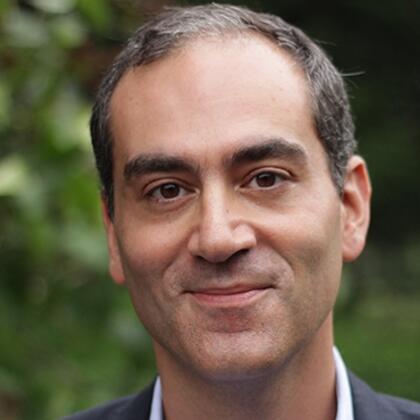
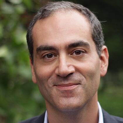
The Neuroscience of Sleep and its Disorders
Share
- Details
- Text
- Audio
- Downloads
- Extra Reading
A good night's sleep is anything but quiet: a myriad of processes occupy our brains, crucial for every aspect of our waking lives. Our increased understanding of the neuroscience of sleep – that sleep may not affect the brain in its entirety – provides a window into the human experiences of sleep deprivation, lucid dreaming, spiritual visitations and a range of clinical sleep disorders, such as insomnia, dream enactment and sleep paralysis.
Download Text
The Neuroscience of Sleep and its Disorders
Prof Guy Leschziner
16th March 2022
In the biomedical sciences, and in the field of neuroscience in particular, experimentation on normal subjects has been fundamental to the increased understanding of the functioning of the human body and brain. However, medicine has long relied upon insights from disease and dysfunction. These constitute Nature’s experiments, where damage or illness, and the consequences thereof, have provided important opportunities to comprehend normal physiological function and structure.
In the world of clinical neuroscience, there are countless examples. Perhaps one of the most famous of these is Phineas Gage, who in 1848 was working in railroad construction in Vermont, USA. While packing explosives into a hole with a heavy tamping iron to blast away rock, he must have sparked the iron against the rock, and ignited the explosive. The tamping iron was driven up with great force, entering Gage’s body under the chin, and exiting the top of his skull on the left. Remarkably, after a brief seizure, Gage sat up, and survived this terrible accident, but he was not left unscathed. Whereas prior to being a civil, God-fearing man, after the accident he was reported to be foul-mouthed, belligerent and vulgar, his personality transformed by the destruction of the left frontal lobe of the brain. Gage’s unfortunate accident provided an early and dramatic illustration of a feature that now represents a concept that is at the core of clinical neurology, that of localization. The central and peripheral nervous system is highly organized, and different functions are located in different regions. In the evaluation of patients with neurological disorders, the first step is to localize the area of damage – the neurological “lesion” describes the anatomical location of damage, injury or dysfunction.
In a clinical setting, disorders of sleep have historically fallen within the remit of respiratory physicians, largely due to the early identification of obstructive sleep apnoea (OSA) – the obstruction of the airway during sleep – as a cause for poor quality sleep and excessive daytime sleepiness. This is despite a wealth of evidence that “sleep is of the brain, by the brain and for the brain” (J Allan Hobson, 2005) – that is, sleep is a process that is controlled and regulated by the brain, and its major functions are to maintain various aspects of the brain, both physiological and psychological. Indeed, many of the sleep disorders that are seen in a clinical setting demonstrate how lesions within the brain give rise to disease and dysfunction with regard to sleep. These lesions are usually less dramatic than Gage’s tamping iron. They are frequently transient, microscopic or functional, but they are lesions nonetheless. Sometimes, the “lesioning” of sleep itself may also provide important insights into the relevance of sleep for the brain itself. In the course of this lecture, I will aim to demonstrate what sleep disorders tell us about ourselves.
Normal Sleep
It is tempting to consider sleep as simply representing an “off” state of the brain, a period during which the brain is quiescent and resting. We have known of course that this is not the case for time immemorial; the psychological experience of dreaming informs us that this is not the case. However, it is only in the last 70 years that we have understood that sleep does not consist of “brain rest”. The development of the electroencephalogram (EEG), which measures electrical activity of cerebral cortex - the surface of the brain – by attaching electrodes to the scalp, has demonstrated that, rather than being an off-state of the brain, sleep represents a dynamic series of states, constantly shifting throughout the night. In fact, in certain stages of sleep, namely rapid eye movement (REM) sleep, on an electrical basis the brain looks very similar to the awake brain, and appears to be highly active. The typical adult cycles through the different stages of sleep roughly every 90 minutes or so, with between four and five cycles of the course of the average night.
However, even this view of sleep, as a dynamic series of brain states, is an over-simplification. Many of the sleep disorders seen in the clinical setting, and indeed normal phenomena that we may all experience, are as a result of sleep not even being a global brain state, meaning that the whole of the brain may not exist in the same stage of sleep or wake. It is this blurring of the margins of various sleep stages that explains many pathologies of sleep.
The Sleep Disorders
Non-REM Parasomnias
Non-REM parasomnias are defined as abnormal behaviours arising from the deepest stages of sleep. The range of behaviours is wide, and commonly features sleeptalking, confusional arousals, night terrors and sleepwalking (Drakatos and Leschziner, 2018). Rarer forms of behaviours include sleep-related eating disorder or sexually-driven events (sexsomnia). Non-REM parasomnias are relatively common in childhood, with approximately 20% exhibiting some form of this condition. Clinically significant non-REM parasomnias are less frequent in adults, seen in approximately 2%.
It seems paradoxical that sufferers can exhibit incredibly complex behaviours such as driving, cooking or even rewiring household electrical items in sleep, and the underlying nature of these events has only better been understood in the last few decades. Imaging of the brains of people in the midst of events, using scans that monitor brain activity, have shown that regions of the brain involved in rational thinking, planning, awareness and memory show activity consistent with persisting deep sleep, whilst other areas responsible for movement, reward and emotions show activity consistent with wake (Bassetti et al, 2000). Studies of electrical activity using electrodes implanted into the brain substance itself show electrical changes that corroborate these findings (Terzaghi et al, 2009). These results imply that, at least in people experiencing non-REM parasomnias, that in addition to sleep not being a single brain state, sleep can also be a local brain state, i.e. affecting different parts of the brain to different degrees simultaneously.
Relevance to Normal Individuals
The phenomenon of “local” rather than “global” sleep has long been recognized in certain species, such as aquatic mammals and certain species of birds. Many of these species are able to sleep in one half of their brain at a time (termed “unihemispheric sleep”), permitting sleep to be achieved while continuing to be able to fly, swim or surface to breathe. Unihemispheric sleep has not been identified in humans, but recent evidence points to humans being able to regulate sleep differentially between the two halves of their brain. When humans are brought into the strange environment of a sleep laboratory for the first time, the dominant hemisphere has been shown to achieve less deep sleep and be more sensitive to environmental stimuli. This normalizes after the first night, suggesting that this may provide an explanation for poor sleep in a strange environment (Tamaki et al, 2016). It is proposed that this may be an evolutionary mechanism for monitoring threat in a novel environment in sleep.
In fact, there is increasing evidence that “local” sleep may exist in full wakefulness, and this may explain why acute sleep deprivation affects cognition and performance. Experimental studies in rats suggest that small areas of the cerebral cortex will repeatedly go silent, and with increasing sleep deprivation these silent periods will become more prolonged and frequent, in parallel with worsening performance at particular tasks (Vyazovskiy et al, 2011). Findings on scalp EEG recordings in humans provide support for this phenomenon occurring in humans (Siclari and Tononi, 2017).
REM sleep is the stage of sleep that most closely correlates with dreaming, particularly dreams of a narrative structure. The hallmark of normal REM sleep is the development of paralysis in all voluntary muscles, with the exception of the muscles that move the eyes (hence the term “REM”).
However, it has long been recognized that certain individuals “act out” their dreams, demonstrating actions that are appropriate to their recall of the content of their dreams. Actions are typically but not invariably aggressive or related to strong emotions. This phenomenon, termed REM sleep behaviour disorder (RBD) is sometimes associated with certain medications, other sleep disorders, or neurological disorders, but principally RBD is a disorder affecting the elderly, men more than women. The underlying cause is thought to relate to a failure of the habitual mechanisms of REM paralysis that are normally mediated by areas with a region of the brain known as the brainstem.
Until recently, RBD was largely considered an “idiopathic” disorder, essentially without any clear cause or connotations. However, in latter years, long-term follow-up of cohorts of people with this disorder have revealed that the vast majority of individuals go on to develop neurodegenerative disorders such as Parkinson’s disease and related conditions, up to 90% after 15 years (Iranzo et al, 2014). Closer study of individuals with RBD show subtle changes in smell, gut and bladder function, the cardiovascular system and cognition, formes frustes of the neurodegenerative disorders associated with RBD.
RBD can be considered another example of local sleep states. While the majority of the brain is in REM sleep, the lack of paralysis that is a feature of wakefulness or non-REM sleep is also present. It is no longer considered an idiopathic condition, and is largely thought of as a prodrome of neurodegeneration, preceding frank Parkinson’s disease or similar disorders by up to 35 years. This provides a significant opportunity for trials of potential disease-modifying drugs in individuals at high risk.
Narcolepsy
Narcolepsy is the archetypal sleep disorder, causing severe sleepiness, sometimes with sudden sleep attacks, hallucinations and/or paralysis as people transition in or out of sleep, and cataplexy – the sudden loss of muscle strength usually precipitated by strong emotions or laughter (Leschziner, 2014). It is relatively rare, affecting about one in 2000 individuals, and onset is often in teenage years. Once narcolepsy develops, it is lifelong.
The origins of narcolepsy were a puzzle until recently. Historically, similarities were identified with aspects of a condition known as encephalitis lethargica, a debilitating condition which occurred in the early 20th century, and has been speculated to be as a result of inflammation of the brain triggered by Spanish flu. Encephalitis lethargica was characterized by significant damage to deep brain areas, including a region called the hypothalamus, and pathologists of the time hypothesed that in narcolepsy, similar areas of the brain were damaged, but that this damage was not visible.
Significant insights into the origins of narcolepsy came from a breeding programme for dogs with familial cataplexy in Stanford in the 1970s. Painstaking genetic work identified that, in dogs at least, mutations in particular genes responsible for structures detecting a specific chemical in the brain, subsequently named hypocretin or orexin. However, such mutations were not found in humans, where the vast majority of narcolepsy is not familial. Subsequent research did though identify that individuals with narcolepsy had absent of deficient levels of this chemical in their spinal fluid, and that in the brains of people with narcolepsy, the roughly 60-80000 nerve cells that produce hypocretin in the hypothalamus, are missing or substantially depleted.
Several strands of evidence point to the immune system as being responsible for the loss of these nerve cells. The strongest pointer is the very high (>98%) correlation between narcolepsy and a particular genetic marker that influences how the immune system responds to environmental or infectious triggers. Narcolepsy is now seen as being caused by the immune system directing an attack against aspects of certain infectious triggers (such as H1N1 swine flu) that look structurally similar to hypocretin, and that these nerve cells are bystander casualties to this immune response in genetically vulnerable individuals.
The hypocretin system has an important role in stabilizing sleep and wake, and as a result of loss of hypocretin, wakefulness becomes unstable (hence sleepiness and sleep attacks), as does REM sleep. The result of flicking in and out of REM sleep directly from or into wakefulness is the experience of aspects of dreaming sleep in full wake. This explains dream mentation in the waking state, which is thought to underlie the hallucinations at sleep onset or offset, and the inappropriate perseveration of the paralysis of REM sleep in wakefulness. Cataplexy is speculated to be a manifestation of REM paralysis inappropriately switched on, although as to why this should be triggered by strong emotion remains a mystery. Some researchers have proposed that strong emotions triggering paralysis are the basis for tonic immobility, the “playing dead” response of some animals to threat, and that in humans this pathway is normally inhibited by hypocretin. Therefore many (but not all) symptoms of cataplexy represent other examples of the blurring of boundaries between different aspects of sleep and wake.
Lucid Dreaming
Lucid dreaming describes the phenomenon of dreaming while being aware that one is in a dream, sometimes with conscious control of the dream narrative. Many normal individuals describe the ability to lucid dream, but it is especially common in individuals with narcolepsy.
Previously considered by some as not a real phenomenon, remarkable work in 2011 clearly demonstrated lucid dreaming to have a neurobiological basis. Lucid dreamers were placed in a scanner which identifies blood flow changes within the brain. They were asked to clench their left and right hand alternately, and the appropriate hand areas of the brain were highlighted. In lucid dreaming, they were asked to dream the same actions, and similar areas of the brain were highlighted (Dresler et al, 2011), thus proving the biological nature of this phenomenon.
In people with narcolepsy, unstable REM sleep provides a ready explanation for lucid dreaming, but in non-narcolepsy lucid dreamers, changes in the connections between different parts of the brain and increased waking-like activity in the frontal areas of the brain have been found. These findings remain controversial, but electrical stimulation of these areas has been shown to increase the lucidity of dreams in normal subjects. It is proposed that, as with non-REM parasomnias, wake-like activity in regions of the brain responsible for awareness is the basis for lucid dreaming, and therefore this might provide insights into the neurobiological basis of consciousness.
Similarly, many individuals with insomnia demonstrate a phenomenon known as paradoxical insomnia. This describes individuals who feel that they are awake while recordings of their brain activity show them to be asleep. More detailed studies of electrical activity suggest that something similar may be at the heart of paradoxical insomnia, that certain regions of the brain do not achieve the same depth of sleep as in normal individuals (Lecci et al, 2020).
Thus far, we have focused on sleep disorders that have a primary neurobiological basis. However, there is much to be learnt even from sleep disorders that have origins elsewhere. Apart from insomnia, the commonest sleep disorder is OSA, whereby anatomical changes within in the airway (often related to genetic factors, lower jaw anatomy or obesity) result in recurrent obstruction of the airway in sleep. These obstructions cause both sleep fragmentation and recurrent drops in blood oxygen at night.
Both insomnia and OSA have been linked to a variety of health outcomes, including cardiovascular disease and Alzheimer’s disease. Why this should be the case is in part explained by the recent discovery of a system of drainage channels within the brain, called the glymphatic system, the role of which is to act as a “waste disposal system”, removing substances from the brain back into the circulation (Xie et al, 2013). It has been shown that sleep, particularly deep non-REM sleep, amplifies the function of the glymphatic system, with a 60% increase in flow in deep sleep. One of the important products removed is a protein called beta-amyloid, the protein deposited in and between brain cells in Alzheimer’s disease. Even a single night of sleep deprivation appears to have a significant impact on levels of amyloid in the spinal fluid and in the brain itself, thus providing a potential mechanism for this link between limited or disrupted sleep and cognitive decline. This effect may be augmented in OSA, where recurrent drops in oxygen may trigger inflammation in the central nervous system, contributing to damage (Polsek et al, 2018).
Conclusions
In conclusion, sleep disorders provide important insights into the nature and function of sleep. Through their study, they show us that sleep is not a single brain state, nor is it a global brain state, and that the view of distinct stages of sleep is an oversimplification. Furthermore, they illustrate the role of the brain in the regulation of sleep, permitting an increased understanding of the circuits and neurotransmitters that control sleep and wake. Finally, through the study of these disorders, we begin to fathom the neurobiological functions of sleep, thus recognizing that sleep is indeed “of the brain, by the brain and for the brain”.
© Professor Guy Leschziner 2022
References and Further Reading
Bassetti C, Vella S, Donati F, Wielepp P, Weder B. SPECT during sleepwalking. Lancet 2000, 356 (9228): 484-5.
Drakatos P, Leschziner G. Diagnosis and management of nonrapid eye movement parasomnias. Curr Opin Pulm Med 2019, 25 (6): 629-635.
Dresler M, Koch SP, Wehrle R, Spoormaker VI, Holsboer F, Steiger A, Saemann PG, Obrig H, Czisch M. Dreamed movement elicits activation in the sensorimotor cortex. Curr Biol 2011, 21 (21): 1833-7.
Hobson JA. Sleep is of the brain, by the brain and for the brain. Nature 2005, 437 (7063): 1254-6.
Iranzo A, Fernandez-Arcos A, Tolosa E, Serradell M, Molineuvo JL, Valldeoriola F, Gelpi E, Vilaseca I, Sanchez-Valle R, Llado A, Gaig C, Santamaria J. Neurodegenerative disorder risk in idiopathic REM sleep behavior disorder: study in 174 patients. PLOS One 2014, 9 (2): e89741.
Lecci S, Cataldi J, Betta M, Bernardi G, Heinzer R, Siclari F. Electroencephalographic changes associated with subjective under- and overestimation of sleep duration. Sleep 2020, 43 (11): zsaa094.
Leschziner G. Narcolepsy: a clinical review. Pract Neurol 2014, 14 (5): 323-31.
Polsek D, Gildeh N, Cash D, Winsky-Sommerer R, Williams SCR, Turkheimer F, Leschziner GD, Morrell MJ, Rosenzweig I. Obstructive sleep apnoea and Alzheimer’s disease: in search of shred pathomechanisms. Neurosci Biobeha Rev 2018, 86: 142-149.
Siclari F, Tononi G. Local aspects of sleep and wakefulness. Curr Opin Neurobiol 2017, 44: 222-227.
Tamaki M, Bang JW, Watanabe T, Sasaki Y. Night watch in one brain hemisphere during sleep associated with the first-night effect in humans. Curr Biol 2016, 26 (9): 1190-1194.
Terzaghi M, Sartori I, Tassi L, Didato G, Rustioni V, LoRusso G, Manni R, Nobili L. Evidence of dissociated arousal states from an intracerebral neurophysiological study. Sleep 2009, 32 (3): 409-12.
Vyazovskiy V, Olcese U, Hanlon EC, Nir Y, Cirelli C, Tononi G. Local sleep in awake rats. Nature 2011, 472 (7344): 443-7.
Xie L, Kang H, Xu Q, Chen MJ, Liao Y, Thiyagarajan M, O’Donnell J, Christensen DJ, Nicholson C, Iliff JJ, Takano T, Deane R, Nedergaard M. Sleep drives metabolite clearance from the adult brain. Science 2013, 342 (6156): 10.1126.
Part of:
This event was on Wed, 16 Mar 2022
Support Gresham
Gresham College has offered an outstanding education to the public free of charge for over 400 years. Today, Gresham College plays an important role in fostering a love of learning and a greater understanding of ourselves and the world around us. Your donation will help to widen our reach and to broaden our audience, allowing more people to benefit from a high-quality education from some of the brightest minds.


 Login
Login


